Muscles
Structure
 |
Actin
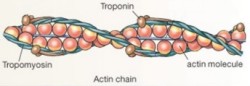 The thin filaments consist of globular actin molecules in two long chains wound around each other and each actin molecule has a binding site for myosin. The protein tropomyosin winds around the thin filaments and covers the myosin binding sites. At regular intervals along the tropomyosin cable sit troponin molecules.
Myosin
The thick filaments consist of bundles of myosin molecules. Each myosin molecule is composed of two long protein chains with a globular head at one end. The myosin head attaches to the binding site on the actin filament. In addition, it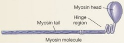 binds ATP, acting as an enzyme to transfer energy from ATP. The energy transfer changes the shape of the myosin head ("cocks" the apparatus).
ContractionThe thin filaments consist of globular actin molecules in two long chains wound around each other and each actin molecule has a binding site for myosin. The protein tropomyosin winds around the thin filaments and covers the myosin binding sites. At regular intervals along the tropomyosin cable sit troponin molecules.Myosin
The thick filaments consist of bundles of myosin molecules. Each myosin molecule is composed of two long protein chains with a globular head at one end. The myosin head attaches to the binding site on the actin filament. In addition, it
binds ATP, acting as an enzyme to transfer energy from ATP. The energy transfer changes the shape of the myosin head ("cocks" the apparatus).When muscle contracts, the globular heads of the thick myosin filaments attach to the binding sites on the thin actin filaments and pull them toward each other. Since the thin filaments are anchored in the Z line, the sliding of the filaments causes each sarcomere - and thus the muscle fibers - to shorten.
Setup
Muscle contraction and relaxation occurs in a cycle that uses and reuses the same components. Initially, the myosin head combines with ATP cocking its head with the energy from ATP. Before the nerve sends a signal to the muscle to contract, the cocked head cannot bind to the thin actin filament since the binding site is blocked by the troponin-tropomyosin complex.
Shortening
The action potential traveling along the nerve cell releases a chemical messenger (acetylcholine) which crosses the neuromuscular junction and depolarizes the muscle fiber membrane in the same way as depolarization occurs in a nerve cell. Upon depolarization, the sarcoplasmic reticulum releases calcium ions. The calcium ions bind to the troponin, changing the shape of the troponin-tropomyosin complex such that the actin binding sites are uncovered. As soon as myosin binds to actin, the cocked head of myosin releases sliding the actin fiber. Once the action potential occurs in the muscle fiber, it takes a few milliseconds for the processes of excitation and coupling to result in an increase in tension. Peak contraction takes place in about 10 ms in fast muscle fibers and about 100 ms in slow fibers.
Relaxation
If a molecule of ATP is present, it binds to myosin. One effect is this binding is to weaken the connection between myosin and actin enough that the myosin head separates from the actin binding site. The second effect is that the enzyme in the head of myosin takes the energy of the ATP molecule and recocks itself.
Clean-up
Once acetylcholine initiates an action potential at the sarcoplasmic reticulum, it is inactivated by an enzyme. The muscle fiber will contract again only if another signal arrives from the nerve. As soon as the calcium ions flood into the muscle cell, the cell rapidly pumps them back to the sacs of the sarcoplasmic reticulum, using energy from ATP, and the troponin-tropomyosin complex returns to its original shape blocking the actin binding sites.
Death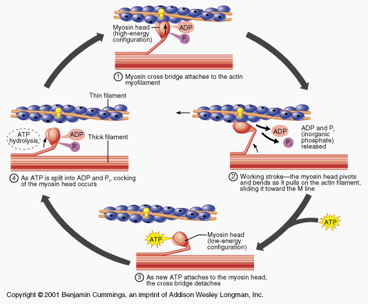 |
Shortening
The action potential traveling along the nerve cell releases a chemical messenger (acetylcholine) which crosses the neuromuscular junction and depolarizes the muscle fiber membrane in the same way as depolarization occurs in a nerve cell. Upon depolarization, the sarcoplasmic reticulum releases calcium ions. The calcium ions bind to the troponin, changing the shape of the troponin-tropomyosin complex such that the actin binding sites are uncovered. As soon as myosin binds to actin, the cocked head of myosin releases sliding the actin fiber. Once the action potential occurs in the muscle fiber, it takes a few milliseconds for the processes of excitation and coupling to result in an increase in tension. Peak contraction takes place in about 10 ms in fast muscle fibers and about 100 ms in slow fibers.
Relaxation
 |
Clean-up
Once acetylcholine initiates an action potential at the sarcoplasmic reticulum, it is inactivated by an enzyme. The muscle fiber will contract again only if another signal arrives from the nerve. As soon as the calcium ions flood into the muscle cell, the cell rapidly pumps them back to the sacs of the sarcoplasmic reticulum, using energy from ATP, and the troponin-tropomyosin complex returns to its original shape blocking the actin binding sites.
When the heart stops beating, neither glucose nor oxygen reaches the cells. Without an energy source or an electron acceptor, the cell cannot pump ions to polarize the membrane, it cannot make ATP, or pump calcium ions out. Without blood flow, calcium leaks into the cells and muscles contract until the cellular supply of ATP is exhausted. Since the muscles cannot relax without ATP to disconnect myosin from actin, the body becomes limp again only when the muscle proteins are digested by lysosomal enzymes during decomposition.
Rigor Mortis
Rigor mortis (stiffening of muscles after death) begins in the smaller muscles of the face and neck in about two hours after death, and proceeds to the feet. The body becomes completely stiff in approximately eight to twelve hours. Bodies remain rigid for approximately eighteen hours, at which time the process begins to reverse itself. In approximately twelve hours the body returns to a flaccid state.
Excerpted and adapted from: Curtis, H., and N. S. Barnes. 1989. Biology. Worth Publishers, Inc., NY., Vander, A., J. Sherman, and Dorothy Luciano. 2001. Human Physiology. The Mechanisms of Body Function. McGraw Hill. Boston., Rigor Mortis and Lividity, What Causes Rigor Mortis?