 |
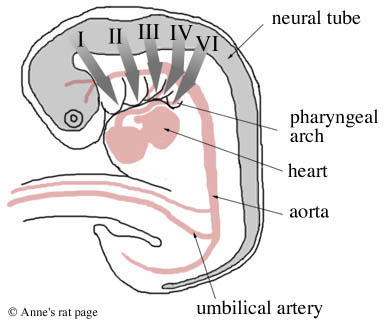
Mammal embryos have five pairs of these pharyngeal arches. The first two pairs give rise to the bones, muscles, and nerves of the ear, jaw and upper neck (arch one becomes the jaws, arch two becomes aspects of the face and ear). The last three pairs of arches give rise to the bones, muscles, and glands (thymus, thyroid) of the neck and the outflow tract of the heart (arch three becomes structures associated with the hyoid and upper pharynx, and arches four and six (arch five disappears) become structures associated with the larynx and lower pharynx; arch four also becomes the aortic arch from the heart; arch six also becomes the pulmonary arteries).
Genetic Control: TCOF1 is one of the many genes involved in pharyngeal arch development. Its protein, Treacle, is expressed most intensely in the first pharyngeal arch during times of critical craniofacial development, including the formation and fusion of the pharyngeal arches with the rest of the face. Treacle is involved in ribosomal RNA production in prefusion neural folds during early embryogenesis.
Another such gene is TBX1, whose gene product has a wide variety of functions. It regulates proper neural crest cell migration in the posterior pharyngeal arches, stabilizes the structural patterns of the middle and inner ear during their development, regulates the onset of the development of jaw and neck muscles, controls the proper patterning of cells in the jaw, supports proper proliferation of cells fated to become part of the cardiac outflow tract, and is required for the formation of the division between the aorta and the pulmonary artery.
Development of the Pharyngeal Arches