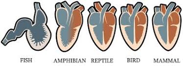
Note that the partial septum in the reptile ventricle becomes a complete divider in birds and mammals.
Key concepts: vertebrate, heart, chamber, double circulation, septum, shunt, atrium, ventricle, vein, artery
Function: The heart is a hollow muscular organ that rythmically contracts and relaxes. During each contraction-relaxation cycle, blood is drawn from the veins into a thin walled collecting chamber, the atrium, and is then passed to a second thick walled chamber, the ventricle, which forceably contracts to distribute the blood to the arteries. Backflow is prevented by one-way valves.
Note that the partial septum in the reptile ventricle becomes a complete divider in birds and mammals.
In the image above, you can see the progressive changes in the heart between ancestral vertebrates, the fishes, and the most derived forms, the birds and mammals. Fish have a simple two chambered heart which is, in essense, just a thickening of a section of the circulatory system, and the blood flows in a single circuit from heart to gills to body and back to the heart. Starting with the amphibians, the first of the vertebrates with lungs, the circulatory system adds a second loop or circuit. This design has the blood flow through the heart twice on each trip around the system, once on the way to the lungs and once on the way back from the lungs, giving it an extra boost. This is called double circulation. In amphibians, with two atria but only a single ventricle, this results in the mixing of deoxygenated and oxygenated blood, but amphibians also gather oxygen through their moist skin, so this inefficiency is not critical. Beginning with the reptiles, a septum or wall develops that partly divides the deoxygenated from the oxygenated blood in the ventricle, and this is important because reptiles, with a watertight skin, rely entirely on their lungs for oxygen. Reptiles also have the unique ability to redirect or shunt blood leaving the heart back through the heart without passing through the body circuit, and to shunt deoxygenated body blood back through the body without going to the lungs. The purpose of this shunt (see the purple vessels in the figure below) is not entirely understood. The former is thought to be a way to prioritize oxygenation of the heart during periods of high exertion, while the latter is believed to be a way to enhance digestion, because of increased acidity of deoxygenated blood due to carbon dioxide buildup. Among the extant reptiles, only the crocodilians have fully extended the septum and have a four-chambered heart, but there is speculation that dinosaurs may have had this innovation as well. Birds and mammals have the same four-chambered design, which has increased efficiency because deoxygenated and oxygenated blood cannot mix.
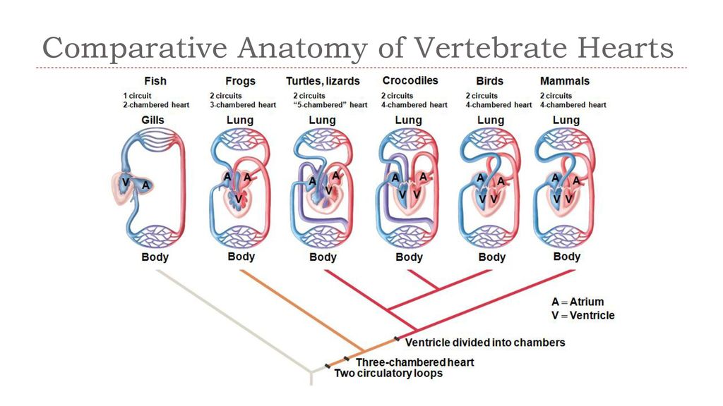
Red blood vessels carry oxygen-rich blood. Purple vessels carry mixed blood. Blue vessels carry deoxygenated blood.