Bacterial Cells

Introduction
Bacteria belong to the Kingdom Prokaryotae (also known as Kingdom Monera) as their cells lack a nucleus. However, it has become increasingly clear that their are two distinct groups of prokaryotes which diverged from each other very early on in the course of evolution. Many researchers are already referring to these groups of bacteria as Domain Eubacteria and Domain Archaea (a domain is a taxonomic ranking above that of Kingdom). A third domain, Eukaryotae, has been proposed which would contain all other organisms with nucleated cells. Bacteria are both ancient and diverse; many have important ecological roles while comparatively few are pathogenic.

Table 1. Key differences between Prokaryotes and Eukaryotes
Feature
Prokaryotic cell
Eukaryotic cell
Nucleus Absent. No nuclear envelope Present with nuclear envelope and nucleolus
Membrane-bound organelles Absent Present. Includes mitochondria, chloroplasts (plants), lysosomes
Chromosome (DNA) Single coiled chromosome in cytoplasm 'nucleoid' region in association with 'histone-like' proteins Multiple linear chromosomes with histone proteins
Cell division (asexual) No true mitotic apparatus. Divide by binary fission or fragmentation Mitosis
Cell wall Eubacteria have a cell wall of peptidoglycan

Archaea have cell walls of pseudomurein

No cell walls in animal cells

Plant cell walls = cellulose

Fungal cell walls = chitin

Ribosomes 70S. Free in cytoplasm 80S. Both free in cytoplasm and attached to rough E.R.

70S in mitochondria and chloroplasts

Cytoskeleton absent present consisting of microtubules and filaments
Flagella when present consist of protein flagellin consist of 9+2 arrangement of microtubules
Cytoplasmic membrane lipids Eubacteria= Fatty acids joined to glycerol by ester linkage

Archaea= Hydrocarbons joined to glycerol by ether linkage

Fatty acids joined to glycerol by ester linkage

Prokaryotic Organisms:

I. Typical Characteristics of prokaryotes

II. Size, Shape and Arrangements III. Structures external to the cell wall. III. Cell wall:
        Gram positive - very thick cell walls. Layers and layers of peptidoglycan. Also contains teichoic acid and lipoteichoic acid.
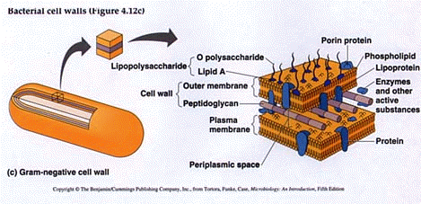
Gram negative - very thin peptidoglycan layer in cell wall. Also has an outer membrane which is considered part of the cell wall. Outer membrane contains porins. The area between the peptidoglycan and cell membrane is called the periplasmic space.
Outer membrane - In Gram negative:
IV. Plasma Membrane

Several names are used: cell membrnane, plasma membrane or cytoplasmic membrane.

MOVEMENT ACROSS THE MEMBRANE:
a) Passive processes (no energy required) b). Active transport IV. CYTOPLASM / CYTOSOL  V. Inclusions
  • Various small particles, crystals and bubbles
  • Polysaccharide (starch, glycogen)
  • Lipids
  • Sulfur
  • Gas
  • VI. RIBOSOMES
  • Particles made up of two subunits (30S and 50S)
  • Subunits are made up of RNA and PROTEIN
  • Proteins are made on ribosomes
  • Procaryotic ribosomes are small (70S) compared with eukaryotic (80S)
  • VII.  NUCLEOID


    PLASMIDS


    VIII. ENDOSPORES
  • These are dormant (not metabolically active) bacterial cells.
  • Endospores are highly resistant to heat, drying, UV radiation, and chemical disinfectants.
  • Bacillus sp. and Clostridium sp. are the most important spore formers in medicine.
  • Germination VS sporulation?
  • Endospores are NOT reproductive structures as only one cell gives rise to one spore.
  • Endospores can be identified with special stains and differentiated from the vegetative cell.
  • During the process of sporulation, a copy of the DNA is pinched off into a separate part of the cell and the cell is protected with a heat-resistant calcified spore coat (dipicolinic acid).
  • The spore contains DNA, a small amount of RNA and little free water. Most metabolic activity shuts down
  • When conditions are favorable there is a rapid shift in metabolic activity and a return to the vegetative form of the cell

  • What is the difference between a vegetative cell and an endospore?