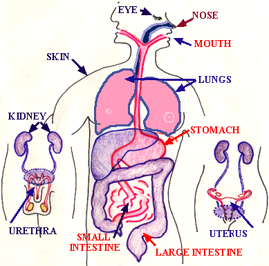
I. Anatomical Barriers and Mechanical Removal

a) Bacterial Antagonism by Normal Flora
Bacteria that are normal body flora keep potentially harmful opportunistic
microorganisms in check and also inhibit the colonization of pathogens
by:
Non-specific host defense mechanisms are distinct from specific host defense mechanisms, which rely upon the specific recognition of the pathogen by lymphocytes. specific immunity is based upon the antibodies made by B-cells and upon the activities and cytokine secretions of T-cells. B-cells and T-cells have receptors, which recognize molecules on the invading organisms. Each b-cell and t-cell has a unique receptor. There are millions of specificities.
A. Phagocytosis
Before we look at phagocytosis in some detail we need to first become
familiar with the various cells in both the bloodstream and in the tissues
and organs of the body that will play a role in body defense.
Defense Cells in the Blood: The Leukocytes
All leukocytes (white blood cells or WBCs) are critical to body defense.
There are normally between 5,000-10,000 leukocytes per cubic millimeter
(mm3) of blood and these can be divided into five major types: neutrophils,
basophils, eosinophils, monocytes, and lymphocytes. The production
of colonies of the different types of leukocytes (leukopoiesis) is induced
by various cytokines known as colony stimulating factors or CSFs.
The five types of leukocytes fall into one of two groups, the polymorphonuclear leukocytes and the mononuclear leukocytes.
Polymorphonuclear leukocytes (granulocytes) have irregular shaped nuclei with several lobes and their cytoplasm is filled with granules containing enzymes and antimicrobial chemicals. They include the following:
Neutrophils
* Neutrophils are the most abundant of the leukocytes, normally accounting
for 54-75% of
the WBCs. An adult typically has 3,000-7,500 neutrophils/mm3
of blood but the number
may increase two- to three-fold during active infections.
* Neutrophils are important phagocytes.
* Their granules contain various agents for killing microbes.
These include lysozyme
(breaks down peptidoglycan), lactoferrin (makes
iron unavailable to bacteria), acid
hydrolase (degrades cellular proteins), and
myeloperoxidase
(catalyzes
reactions that
produce lethal oxidants, including hypochlorous
acid, free chlorine, hydrogen peroxide,
and hydroxyl radicals). These agents kill
microbes intracellularly during phagocytosis
but are also often released extracellularly where
they kill not only microbes but also
surrounding cells and tissue, as will be discussed
later under phagocytosis.
* They release the enzyme kallikrein which catalyzes the generation
of bradykinins.
Bradykinins promote inflammation by causing
vasodilation, increasing vascular
permeability, and increasing mucous production.
They are also chemotactic for leukocytes
and stimulate pain.
* They release enzymes which catalyze the synthesis of prostaglandins
from arachidonic
acid in cell membranes. Certain prostaglandins
promote
inflammation by causing
vasodilation and promoting capillary permeability.
They also cause constriction of
smooth muscles, enhance pain, and induce fever.
Eosinophils
Eosinophils normally comprise 1-4% of the WBCs (50-400/mm3 of blood).
* Their granules contain destructive enzymes for killing infectious
organisms. These
enzymes include acid phosphatase, peroxidases,
and proteinases.
* The substances they release defend primarily against fungi, protozoa,
and parasitic worms,
pathogens that are too big to be consumed by phagocytosis.
Basophils
* Basophils normally make up 0-1% of the WBCs (25-100/mm3 of blood).
* Basophils release histamine which promotes inflammation
by causing vasodilation,
increasing capillary permeability, and increasing
mucous production.
Mononuclear leukocytes (agranulocytes) have compact nuclei and have no visible cytoplasmic granules. The following are agranulocytes:
Monocytes
* Monocytes normally make up 2-8% of the WBCs (100-500/mm3 of
blood).
* Monocytes are important phagocytes.
* Monocytes become macrophages and dendritic cells when they
leave the blood and
enter tissues. Macrophages and dendritic cells are
very important in phagocytosis and
aid in the immune responses (see below).
They produce a variety of cytokines that
play numerous roles in body defense.
Lymphocytes
* Lymphocytes normally represent 25-40% of the WBCs (1,500-4,500/mm3
of blood).
* Lymphocytes mediate the specific immune responses
* Only a small proportion of the body's lymphocytes are found in the
blood. The majority
are found in lymphoid tissue.
* Lymphocytes circulate back and forth between the blood and the
lymphoid
system of the body.
* There are 2 major populations of lymphocytes:
Defense Cells in the Tissue: Macrophages, Dendritic Cells, and Mast Cells
Macrophages
When monocytes leave the blood and enter the tissue, they become activated
and differentiate into macrophages. Those that have recently left the blood
during inflammation and move to the site of infection through positive
chemotaxis are sometimes referred to as wandering macrophages.
In addition, the body has macrophages already stationed throughout the tissues and organs of the body. These are sometimes referred to as fixed macrophages.
Many fixed macrophages are part of the lymphoreticular (reticuloendothelial) system. They are found supported by reticular fibers (along with B-lymphocytes and T-lymphocytes) in lymph nodules, lymph nodes, and the spleen where they filter out and phagocytose foreign matter such as microbes.
Similar cells derived from stem cells, monocytes, or macrophages are also found in the liver (Kupffer cells), the kidneys (mesangial cells), the brain (microglia), the bones (osteoclasts), and the lungs (alveolar macrophages).
Macrophages actually have a number of very important functions in body defense including:
Dendritic cells
Dendritic cells are cells with numerous pseudopodia-like projections
and are located in the follicles of the lymphoid tissue (follicular dendritic
cells) as well as the connective tissue of the skin and mucous membranes
(Langerhans' cells). They are derived from bone marrow progenitor cells
and from monocytes.
Dendritic cells function in processing antigens, presenting those antigens to T-lymphocytes, and producing cytokines similar to the macrophages mentioned above. Dendritic cells are considered to be the most potent antigen-presenting cells (APCs) in the body.
Mast cells
Mast cells, found throughout the connective tissue of the skin
and mucous membranes, carry out the same functions as basophils. They release
histamine
which promotes inflammation by causing vasodilation, increasing
capillary permeability, and increasing mucous production. Mast cells are
the cells that usually first initiate the inflammatory response (discussed
later in this unit).
An Overview of Phagocytic Defense
* Infection or tissue injury stimulates cells such as mast cells and
basophils to release
vasodilators to initiate the inflammatory response.
As a result of vasodilation and
increased capillary permeability, phagocytic white
blood cells (neutrophils,
monocytes/macrophages, eosinophils) and other white
blood cells enter the tissue
around the injured site and are chemotactically
attracted to the area of infection. In other
words, inflammation allows phagocytes to enter
the tissue and go to the site of
infection.
Neutrophils are the first to appear and are later
replaced by macrophage.
* Lymph nodules are unencapsulated masses of lymphoid tissue
containing lymphocytes
and macrophages. They are located in the respiratory
tract, the liver, and the
gastrointestinal tract and are collectively referred
to as mucosa-associated lymphoid
tissue or MALT. Examples include the adenoids
and tonsils
in the respiratory tract
and the Peyer's patches on the small intestines.
Organisms entering these systems can be
phagocytosed by fixed macrophages anddendritic
cells and presented to
lymphocytes to initiate the immune responses.
* Tissue fluid (plasma which has left the blood vessels and entered
body tissues and
organs) picks up microbes and then enters the lymph
vessels as lymph. Lymph vessels
carry the lymph to regional lymph nodes.
Lymph nodes contain many reticular fibers
that support the fixed macrophages and dendritic
cells as well as everchanging
populations of circulating B-lymphocytes and T-lymphocytes.
Microbes picked up by
the lymph vessels are filtered outand
phagocytosed in the lymph nodes by these
fixed macrophages and dendritic cells
and presented to the circulating
T-lymphocytes to initiate immune responses. The
lymph eventually enters the circulatory
system at the heart to maintain the fluid volume
of the circulation.
* The spleen contains many reticular fibers that support fixed macrophages
and dendritic
cells as well as everchanging populations of circulating
B-lymphocytes and
T-lymphocytes. Blood carries microorganisms to the
spleen
where
they are filtered out
and phagocytosed by the fixed macrophages
and dendritic cells and presented to
the circulating T-lymphocytes to initiate immune
responses.
* As mentioned above under fixed macrophages, there are also specialized
macrophages
and dendritic cells located in the brain (microglia),
lungs (alveolar macrophages), liver
(Kupffer cells), kidneys (mesangial cells),
bones (osteoclasts), and skin and mucous
membranes (Langerhans' cells).
Steps in Phagocytosis
a) Activation
Resting phagocytes are activated by inflammatory mediators such
as bacterial products, complement proteins, proinflammatory cytokines,
and prostaglandins. As a result, the phagocytes produce surface glycoprotein
receptors that increase their ability to adhere to surfaces and
recognize microbes. They also exhibit increased metabolic and microbicidal
activity (production of ATPs, lysosomal enzymes, lethal oxidants, etc.).
b) Chemotaxis(for wandering macrophages and neutrophils)
Chemotaxis is the movement of phagocytes toward an increasing concentration
of some attractant such as bacterial factors (bacterial proteins, capsules,
cell wall fragments, endotoxin), complement components (C3a, C5a, C5b67),
chemokines (chemotactic cytokines such as interleukin-8 secreted by various
cells), fibrin split products, kinins, and phospholipids released by injured
host cells.
Some microbes, such as the influenza A viruses, Mycobacterium tuberculosis, blood invasive strains of Neisseria gonorrhoeae, and Bordetella pertussis have been shown to block chemotaxis.
c) Attachment
Attachment of microorganisms is necessary for ingestion and may be:
* unenhanced - non-specific attachment to a variety of microbes
by means of glycoprotein
receptors on the surface of the phagocytes; or
* enhanced - attachment by way of the antibodies IgG and IgA
or the complement protein
C3b. Molecules such as IgG, IgA and C3b which promote
enhanced attachment are
called opsonins and the process is called
opsonization.
Enhanced attachment is much
more specific and efficient than unenhanced.
* Organisms with capsules, such as Streptococcus pneumoniae, Neisseria
meningitidis,
and Hemophilus influenzae, may initially
block
attachment of microorganisms; the
exotoxin protein A produced by Staphylococcus
aureus blocks opsonization with IgG.
d) Ingestion
After attachment, the plasma membrane of the phagocyte invaginates,
by
means of the contractile proteins actin and myosin on the inner membrane
surface pulling against rigid microtubules in the cytoplasm, and
pinches
off. This places the ingested organism in a membranous sac called a
phagosome.
Some bacteria, such as pathogenic Yersinia, secrete proteins that depolymerize actin and prevent phagosome formation.
e) Destruction
Phagocytes contain membranous sacs called lysosomes(produced
by the Golgi apparatus) which contain various hydrolytic enzymes and
microbicidal systems. The lysosomes fuse with the phagosomes
and
the microorganisms are killed and digested.
Some bacteria are more resistant to phagocytic destruction once engulfed.
If the phagocyte is overwhelmed with microorganisms, the phagocyte will empty the contents of its lysosomes by a process called degranulation in order to kill the microorganisms or cell extracellularly. These released lysosomal contents, however, also kill surrounding host cells and tissue. Most tissue destruction associated with infections is a result of this process. This is discussed more later under chronic inflammation.
The phagocyte will also empty the contents of its lysosomes for extracellular killing if the cell to which the phagocyte adheres is too large to be engulfed.
There are 2 killing systems in neutrophils and macrophages:
the oxygen-dependent system and the oxygen-independent system.
1) The oxygen-dependent system
In addition to phagocytes using this oxygen-dependant system to
kill microbes intracellularly, neutrophils also routinely release
these oxidizing agents, as well as acid hydrolases, for the purpose of
killing microbes extracellularly. These agents, however, also wind
up killing the neutrophils themselves as well as some surrounding body
cells and tissues as mentioned above.
2) Oxygen-independent system
(2.) INFLAMMATION
This is a general response mounted by the body to virtually any insult.
(If
you recall your own bodies response to a recent cut, puncture wound, or
burn you will be able to appreciate each of the active components of the
inflammatory response.)
i) Reddening --> ii) Swelling ---> iii) Heat --->
iv)
Pain
· A complex reaction triggered by
any damage to the body. Can be provoked by infectious
agents, physical agents, and by certain immune
pathways.
· Symptoms include: pain, redness,
swelling, heat and loss of function.
· Purpose is to destroy the invader,
limit damage and repair the damage.
· There are a number of biochemical
mediators including histamine, prostaglandins,
leukotrienes, complement and kinin.
· These promote vasodilation
and increased vascular permeability as well as phagocyte
and lymphocyte chemotaxis and activation.
· Eventually cytokines and
hormones
stimulate tissue regeneration.
(3.) FEVER
Activated macrophages release interleukin-1 (IL-1). This cytokine
resets the hypothalamus thermostat and the body temperature increases.
Higher temperature may increase the metabolic rate of white cells as well
as slow down the growth of some pathogens. Fever stimulates the release
of transferin an iron binding protein.
(4.) COMPLEMENT ACTIVATION
· Function of activated complement:
a.) destroys cells, b.) stimulates inflammation,
c.) stimulates chemotaxis d) estimulates
phagocytosis.
· Complement is activated
by two different cascades:
The classical pathway and the alternative
pathway
Both pathways activate C3 to form C3a
(inflammatory
mediator) and C3b (opsonin and
enzyme which then activates C5)
· C5 is broken down by C3b
to form C5a (inflammatory mediator and leukocyte attractor)
and C5b (activates C6, C7, C8 and C9 to form
the membrane attack complex (MAC)
which forms the transmembrane channel in the target
cell and leads to cytolysis).