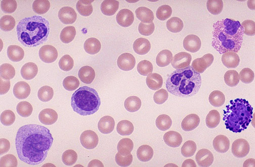
GRANULOCYTES: NEUTROPHIL - EOSINOPHIL - BASOPHIL
AGRANULOCYTES: LYMPHOCYTE - MONOCYTE
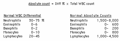
VALUES FOR BLOOD CELLS
NEUTROPHIL
GRANULOCYTES: NEUTROPHIL - EOSINOPHIL - BASOPHIL
AGRANULOCYTES: LYMPHOCYTE - MONOCYTE
VALUES FOR BLOOD CELLS
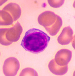
NEUTROPHIL
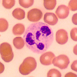
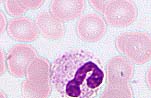
This granulocyte has very tiny light staining granules (the granules
are very difficult to see). The nucleus is frequently multi-lobed with
lobes connected by thin strands of nuclear material. These cells are capable
of phagocytizing foreign cells, toxins, and viruses.
When taking a Differential WBC Count of normal blood, this type of
cell would be the most numerous. Normally, neutrophils account for 50-70%
of all leukocytes. If the count exceeds this amount, the cause is usually
due to an acute infection such as appendicitis, smallpox or rheumatic fever.
If the count is considerably less, it may be due to a viral infection such
as influenza, hepatitis, or rubella.
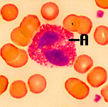
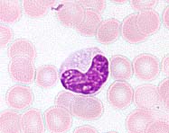
This granulocyte has large granules (A) which are acidophilic and appear
pink (or red) in a stained preparation. This micrograph was color enhanced
to illustrate this feature. The nucleus often has two lobes connected by
a band of nuclear material. (Does it looks like a telephone receiver?)
The granules contain digestive enzymes that are particularly effective
against parasitic worms in their larval form. These cells also phagocytize
antigen - antibody complexes.
These cells account for less than 5% of the WBC's. Increases beyond this amount may be due to parasitic diseases, bronchial asthma or hay fever. Eosinopenia may occur when the body is severely stressed.
In a Differential WBC Count we rarely see these as they represent less
than 1% of all leukocytes. If the count showed an abnormally high number
of these cells, hemolytic anemia or chicken pox may be the cause.
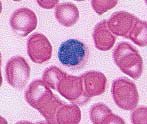
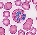
The lymphocyte is an agranular cell with very clear cytoplasm which
stains pale blue. Its nucleus is very large for the size of the cell and
stains dark purple. (Notice that the nucleus almost fills the cell leaving
a very thin rim of cytoplasm.) This cell is much smaller than the three
granulocytes (which are all about the same size). These cells play an important
role in our immune response. The T-lymphocytes act against virus infected
cells and tumor cells. The B-lymphocytes produce antibodies.
This is the second most numerous leukocyte, accounting for 25-35% of
the cells counted in a Differential WBC Count. When the number of these
cells exceeds the normal amount, one would suspect infectious mononucleosis
or a chronic infection. Patients with AIDS keep a careful watch on their
T-cell level, an indicator of the AIDS virus' activity.
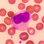
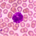
This cell is the largest of the leukocytes and is agranular. The nucleus is most often "U" or kidney bean shaped; the cytoplasm is abundant and light blue (more blue than this micrograph illustrates). These cells leave the blood stream (diapedesis) to become macrophages. As a monocyte or macrophage, these cells are phagocytic and defend the body against viruses and bacteria.
These cells account for 3-9% of all leukocytes. In people with malaria,
endocarditis, typhoid fever, and Rocky Mountain spotted fever, monocytes
increase in number.
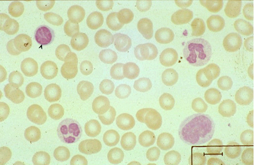
The background cells in this micrograph are erythrocytes (red blood
cells). These cells are non-nucleated, biconcave discs that are filled
with hemoglobin. The primary function of these cells is to carry oxygen
from the lungs to the body cells.
Woman usually have 4-5 million erythrocytes per cubic millimeter of
blood, men have 5-6 million. If this number is considerably higher, polycythemia
may be the cause. If the number is considerably less, the person has anemia.
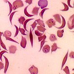
Sickle cell anemia is an inherited condition which results in some
erythrocytes being malformed. The gene for this condition causes the hemoglobin
to be incorrectly formed, which in turn causes some erythrocytes to take
on a crescent shape. These cells are not able to carry adequate amounts
of oxygen to cells.
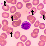
Platelets, which are cell fragments, are seen next to the "t's" above.
(Many of the other micrographs on this page contain them as well.) Platelets
are important for proper blood clotting.
Each cubic millimeter of blood should contain 250,000 to 500,000 of
these. If the number is too high, spontaneous clotting may occur. If the
number is too low, clotting may not occur when necessary.
Do you know what types of cells are these?